Advanced Equine Lameness Evaluation: A Deep Dive
By Claire Halden
Beyond the routine check, understanding nuanced equine lameness requires a comprehensive approach. Discover advanced diagnostics and management strategies for your horse.
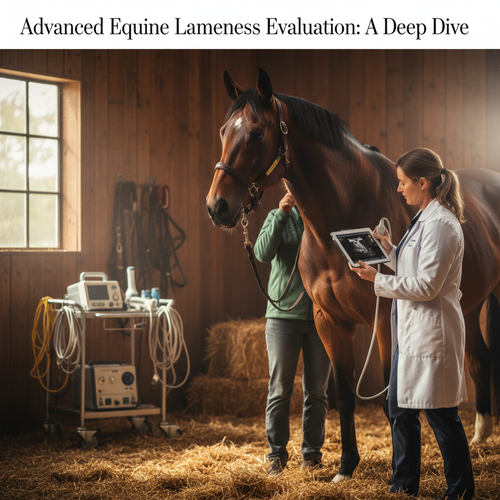
More than 1.5 million horses compete annually in the United States, and a staggering 60-70% of performance horses will experience some degree of lameness within their career, according to various studies cited by the American Association of Equine Practitioners (AAEP). While a basic veterinary examination can identify overt issues, truly understanding the complexities of equine lameness often requires a deeper, more systematic approach. This article delves into advanced evaluation techniques, helping horse owners, trainers, and barn managers recognize subtle signs and pursue comprehensive diagnostic pathways.
Understanding the Nuances of Equine Lameness
Lameness is defined as any alteration in a horse's gait or stance, often due to pain, injury, or a mechanical defect in the musculoskeletal system. It's not a diagnosis in itself, but rather a clinical sign pointing to an underlying problem. The challenge lies in pinpointing the exact source and nature of that problem.
The Limitations of Basic Evaluation
A routine veterinary check typically involves observation at rest, palpation, flexion tests, and observation in hand at a walk and trot. While foundational, this often falls short for subtle lameness cases. For example, a low-grade hind limb lameness might only manifest under saddle or at higher speeds, making in-hand evaluations insufficient. Furthermore, compensatory lameness (where one limb hurts, causing the horse to shift weight, leading to pain in another) can easily mask the primary issue.
When to Suspect Deeper Issues
Beyond overt head-bobbing or severe toe-dragging, watch for:
- Subtle asymmetry: Uneven muscle development, particularly in the hindquarters or shoulder.
- Performance decline: Reluctance to engage, difficulty with specific leads, loss of impulsion, or resistance under saddle.
- Behavioral changes: Increased irritability, anxiety, or resistance to handling/grooming in specific areas.
- Unusual wear patterns: On hooves or shoes that don't correlate with typical conformation.
- Chronic issues: Recurring "off" days despite rest or conventional treatments.
Advanced Diagnostic Techniques: Beyond Visual Assessment
Once basic checks suggest a deeper problem, advanced diagnostics become crucial. These methods provide objective data and detailed imaging, helping veterinarians pinpoint the precise location and nature of the injury.
1. Lameness Locator Systems (Quantitative Gait Analysis)
These systems, like those offered by Equinosis or Lameness Locator, use wireless inertial sensors placed on the horse (head, sacrum, and limb(s)) to objectively measure subtle asymmetries in movement. The sensors record vertical motion, and proprietary algorithms analyze the data to identify and quantify lameness, often with a sensitivity far exceeding the human eye.
- How it works: Sensors transmit data to a computer, which generates a report showing the degree and limb of lameness. It can detect asymmetries as small as 1-2mm that are imperceptible to observers.
- Benefits: Objectivity, repeatability, ability to detect subtle lameness, and track response to treatment. It's especially valuable for multi-limb lameness or when trying to isolate which limb is primary.
- Cost: Typically ranges from $250 - $600 per evaluation, depending on region and clinic. Often combined with a regular lameness exam fee.
2. Diagnostic Anesthesia (Nerve Blocks and Joint Blocks)
This remains a cornerstone of lameness diagnosis. By systematically desensitizing areas of the limb using local anesthetics, veterinarians can isolate the source of pain.
- Nerve Blocks: Anesthetic is injected near specific nerves, numbing the region they supply. Starting distally (e.g., foot) and working proximally (e.g., knee/hock), the horse is trotted after each block. When the lameness improves or resolves, the pain source is identified within the blocked area.
- Joint Blocks: Anesthetic is injected directly into a joint or synovial structure (e.g., tendon sheath, bursa). This directly targets potential pain originating within that specific structure.
- Considerations: Requires meticulous technique to avoid false positives/negatives. Effects are temporary. Potential for temporary weakness or stumbling in blocked limb.
3. Advanced Imaging Modalities
Once the region of interest is identified (often via blocks or gait analysis), advanced imaging provides a detailed view of the underlying structures.
a. Radiography (X-rays)
Still fundamental, radiographs are excellent for evaluating bone structure, identifying fractures, arthritis, bone cysts, and certain developmental abnormalities (e.g., osteochondrosis).
- Limitations: Poor at visualizing soft tissues (tendons, ligaments, cartilage).
- Cost: Varies by number of views and region, typically $150 - $400 per area.
b. Ultrasonography (Ultrasound)
Excellent for soft tissue assessment, ultrasound visualizes tendons, ligaments, joint capsules, menisci, and fluid accumulations. It can detect tears, inflammation, effusions, and changes in tissue texture.
- Benefits: Real-time imaging, portable, no radiation. Crucial for diagnosing suspensory ligament injuries, bowed tendons, and collateral ligament issues.
- Limitations: Operator-dependent, poor penetration of bone. Air can obscure images.
- Cost: $200 - $500 per area, depending on complexity and location.
c. Magnetic Resonance Imaging (MRI)
Often considered the gold standard for soft tissue and bone detail, MRI provides exquisite cross-sectional images without radiation. It's particularly valuable for complex foot lameness, bone bruises, early stress fractures, and subtle soft tissue lesions not visible on X-ray or ultrasound.
- Benefits: Unparalleled detail for both bone and soft tissues, especially within the hoof capsule or complex joint structures.
- Types: High-field (requires general anesthesia, higher image quality) and low-field (standing, less image quality but safer for certain horses).
- Limitations: High cost, limited availability, typically requires referral to specialized clinics. High-field MRI necessitates general anesthesia, carrying inherent risks.
- Cost: $1,500 - $3,000+ per region, depending on type (standing vs. general anesthesia) and clinic.
d. Computed Tomography (CT Scan)
CT uses X-rays to create detailed cross-sectional images, superior to conventional X-rays for complex bony structures like the skull, vertebrae, and distal limb. It's excellent for assessing fracture configurations, bone infections (osteomyelitis), and dental disease.
- Benefits: Excellent for complex bony anatomy. Faster than MRI for many applications. Can be performed standing or under anesthesia.
- Limitations: Limited soft tissue detail compared to MRI. Involves radiation.
- Cost: $1,000 - $2,500+ per region.
e. Scintigraphy (Bone Scan)
This nuclear medicine technique uses a radioactive tracer (technetium-99m) injected intravenously. The tracer accumulates in areas of increased bone metabolism (e.g., inflammation, remodeling, stress fractures, active arthritis). A special camera detects the emitted radiation, highlighting "hot spots" where bone turnover is elevated.
- Benefits: Identifies active bone lesions anywhere in the body, useful for diffuse or challenging to localize lameness. Can detect problems before they are visible on X-ray.
- Limitations: Not specific; a "hot spot" indicates activity but not the precise pathology. Horse must be isolated for 24-48 hours post-injection due to radioactivity.
- Cost: $1,000 - $1,800 for a full body scan.
Practical Management and Prognosis
Once a diagnosis is made, the focus shifts to treatment and management. This might involve anything from targeted rest and medication to advanced regenerative therapies or surgery.
Importance of Holistic Management
Effective lameness management extends beyond the immediate injury. Consider:
- Footing: Optimal arena footing can significantly reduce concussion and strain. Consult with experts on appropriate depths and materials.
- Farriery: Corrective or therapeutic shoeing is paramount. A skilled farrier working in conjunction with your veterinarian can dramatically impact recovery and prevent recurrence. Uneven hoof growth or improper angles can exacerbate lameness, as discussed in publications like The Horse magazine.
- Nutrition: Adequate nutrition supports tissue repair and overall health. Protein, minerals (calcium, phosphorus, copper, zinc), and vitamins (A, D, E) are vital. Tools like the Horse Feed Calculator can help ensure balanced diets, while the Horse Weight Calculator helps maintain ideal body condition to minimize joint stress.
- Exercise Program: Carefully managed rehabilitation programs are critical for strengthening tissues and regaining function. This often involves controlled hand walking, specific exercises, and gradual return to work.
- Environment: Stable hygiene, access to turnout, and stress reduction all play roles in recovery.
Regional Cost Variations
It's important to note that diagnostic and treatment costs can vary significantly by region. For instance, advanced imaging in a major equestrian hub like Lexington, KY, or Ocala, FL, might have different pricing structures compared to more rural areas. Always obtain estimates from your veterinarian. The USDA's regional economic reports often highlight these variances across agricultural services, including equine veterinary care.
Conclusion
Evaluating equine lameness effectively requires moving beyond the obvious. A comprehensive approach integrating objective gait analysis, precise diagnostic blocks, and advanced imaging technologies allows for accurate diagnosis and targeted treatment plans. By understanding these tools and working closely with your veterinarian and other equine professionals, you can provide the best possible care, extend your horse's athletic career, and ensure their long-term comfort and well-being.
Frequently Asked Questions (FAQ)
Q: How often should I have my horse checked for lameness, even if they seem fine?
A: While there's no universal rule, a routine veterinary exam at least once or twice a year is advisable. For performance horses, pre-purchase exams, pre-season check-ups, and post-season evaluations can catch subtle issues early. Some owners opt for objective lameness evaluations (e.g., with a Lameness Locator) annually to establish a baseline.
Q: Can nutrition really impact lameness?
A: Absolutely. Deficiencies or imbalances in key nutrients, especially minerals like copper and zinc, can affect bone and cartilage development and repair. Overfeeding, leading to obesity, places undue stress on joints. Conversely, adequate protein and energy are essential for muscle strength and tissue healing. Discuss your horse's diet with your vet and an equine nutritionist.
Q: What is the biggest mistake owners make when dealing with lameness?
A: One of the biggest mistakes is failing to pursue a definitive diagnosis, instead opting for symptomatic treatment or prolonged rest without understanding the underlying issue. This can lead to chronic problems, compensatory lameness, and ultimately, a poorer prognosis. Early and accurate diagnosis is key.
Q: Is MRI always necessary for lameness diagnosis?
A: No, not always. MRI is indicated when other diagnostics (blocks, X-rays, ultrasound) have narrowed down a region but haven't pinpointed the exact soft tissue or bony lesion, especially in complex areas like the hoof. For many straightforward lameness cases, less expensive and invasive methods are sufficient.
This article is general information, not veterinary advice. Talk to your veterinarian about your horse.